


Triple Marker with Graph (2nd Trimester)
Also referred as
Triple Marker Pregnancy Test (Graph)
Maternal Screening Test

For women
Earliest reports in

48 hours

Contains

3 tests
Test price:
₹2609
10% off
Get it at ₹2348 with coupon
Apply coupon 1MGNEWG during checkout


More offers available

Know more about this test

The Triple Marker with Graph (2nd Trimester) test is a prenatal screening test typically conducted between 14-22 weeks of gestation, ideally between 15-20 weeks (the second trimester) of pregnancy. It analyzes three key biochemical markers in the blood: Alpha-fetoprotein (AFP), Human chorionic gonadotropin (HCG), and Estriol unconjugated (uE3). Combined with ultrasound findings, this evaluation calculates the risk of certain chromosomal abnormalities and neural tube defects in the developing fetus.


Samples required
Blood

Find out
Why is this test booked?

Preparation for this test

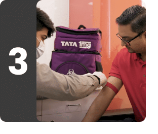
Sample Collection
Who will collect your samples?
Conducted by
Tata 1mg Labs


Accredited
labs
Highly skilled
Phlebos
Verified
reports
Who will collect your samples?
Tata 1mg certified phlebotomists
Know more about lab
Test Information is reviewed by: Dr. Swati Gupta (Anand), MD Lab Medicine, MBBS and written by Dr. Lipika Khurana, PGDHHM, BDS . The test details are for information purpose only. Consult a doctor before taking any test. Last updated on: 2nd Jul 2026
Did you find information useful ?
Book a Pan-Fungal PCR test at home near me
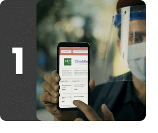
Easy online booking
Search for tests and packages, book your preferred time slot and enter your address for seamless at-home lab tests.
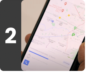
Live tracking of phlebotomist
Track our trained phlebotomist's real time location for seamless sample collection.
Safe Sample Collection
Our phlebotomists follow strict safety protocols to collect samples safely at home and on time.

Sample received at lab
Samples are transported securely to our accredited labs with world-class machines for testing by qualified experts.
Quick, Doctor-Verified Reports
Get doctor-approved reports via email and WhatsApp, with options for doctor follow-ups and AI insights.


References
- Conde-Agudelo A, Kafury-Goeta AC. Triple-marker test as screening for Down syndrome: a meta-analysis. 1998. In: Database of Abstracts of Reviews of Effects (DARE): Quality-assessed Reviews [Internet]. York (UK): Centre for Reviews and Dissemination (UK); 1995-. Available from:

- Genetic Alliance; District of Columbia Department of Health. Understanding Genetics: A District of Columbia Guide for Patients and Health Professionals. Washington (DC): Genetic Alliance; 2010 Feb 17. Appendix F, Maternal Serum Marker Screening. Available from:
- Driscoll DA, Gross SJ; Professional Practice Guidelines Committee. Screening for fetal aneuploidy and neural tube defects. Genet Med. 2009 Nov;11(11):818-21. [Accessed 02 May 2024]. Available from:
- Greene ND, Copp AJ. Neural tube defects. Annu Rev Neurosci. 2014;37:221-42. [Accessed 02 May 2024]. Available from:
- Screening for Down's syndrome, Edwards' syndrome and Patau's syndrome [Internet]. NHS; 19 April 2021 [Accessed 02 May 2024]. Available from:
- Sablok A, Sharma A, Ahmed CS, Kaul A. Performance of second-trimester maternal biochemistry screening (quadruple test vs. triple test) for trisomy 21: An Indian experience. Indian J Med Res. 2021 May;154(5):716-722. [Accessed 02 May 2024]. Available from:
- Gordon S, Umandap C, Langaker MD. Prenatal Genetic Screening. [Updated 2023 Jan 16]. In: StatPearls [Internet]. Treasure Island (FL): StatPearls Publishing; 2023 Jan-. Available from:
- Commit to Healthy Choices to Help Prevent Birth Defects [Internet]. CDC; 28 June 2023 [Accessed 02 May 2024]. Available from:
- Institute of Medicine (US) Committee on Improving Birth Outcomes; Bale JR, Stoll BJ, Lucas AO, editors. Reducing Birth Defects: Meeting the Challenge in the Developing World. Washington (DC): National Academies Press (US); 2003. 3, Interventions to Reduce the Impact of Birth Defects. [Accessed 02 May 2024]. Available from:

Recommended for women
This package is designed with women's health considerations in mind, offering targeted assessments to address unique wellness needs and potential risks.
Contains 3 tests
Report delivery
Standard time
48 hrs
For slots after 11 AM, report will be delivered in 72 hours.
Samples required
Blood
1 vial required
Our phlebotomist will draw a blood sample, typically from a vein in your inner elbow.
Preparations
1
Provide maternal Date of birth (dd/mm/yy); Date of the first day of the last menstrual period (LMP), Ultrasound; Number of Fetuses (Single/Twins); Diabetic status and Body Weight in Kg, IVF, Smoking & Previous history of Trisomy 21 pregnancy at the time of sample collection.
2
A duly filled Maternal Serum Screen requisition form (Annexure - CR/02) is mandatory. Valid between 14-22 weeks gestation (Ideal 15-20 weeks). USG report is required.
Why is this test booked?
1
The Triple Marker with Graph (2nd Trimester) test is done:
2
As a part of standard screening between 14-22 weeks (ideal 15-20 weeks) of pregnancy.
3
To assess the risk of Down Syndrome (Trisomy 21) in the developing baby.
4
To assess the risk of Edwards Syndrome (Trisomy 18) in the developing baby.
5
To assess the risk of neural tube defects like spina bifida in the developing baby.
Coupon details
Get it at ₹2348 with coupon
Get upto 15% off on your lab tests
Maximum discount: ₹1500
1MGNEWG
10% off
₹999
13% off
₹3000
15% off
₹5000
Available Offers
Get additional 15% off
Get 15% off on your first order of lab tests
Maximum discount: ₹300
1MGNEW
Get additional upto 15% off
Get upto 15% off on your lab tests
Maximum discount: ₹1500
1MGNEWG
Upto ₹650 off on all lab tests
Get upto ₹650 off on all lab tests
Maximum discount: ₹650
1MGSUPER
Upto Rs 1500 off on all lab tests
Get upto ₹1500 off on all lab tests
Maximum discount: ₹1500
1MGSUPERMAX
Flat 20% off on all lab tests
Get flat 20% off on all lab tests
Maximum discount: ₹2500
1MGMAX20
Who's behind your sample collection?


MEET YOUR PHLEBO

Certified & experienced professionals
Tata 1mg phlebotomists are DMLT / B.Sc MLT certified and have 1+ years of experience

Best in-class collections
Tata 1mg phlebotomists are trained for painless, single-prick hygienic sample collection

Comprehensive expertise
Other than sample collection, our phlebos are also skilled in first aid, ECG & BP monitoring
WHAT OUR CUSTOMERS ARE SAYING
Pain free collection
~ Savitha S
Very good conduct of collector
~ Lakshmi E
Smooth and quick.
~ Sarabjeet singh
Good behavior
~ Ranjeet Singh
Better service
~ Manisha Kobnak
Very good
~ SANDIP CHHAGAN PATIL 7030019951
Good
~ Dhandapani Kavitha
Ok
~ Aatamjeet Singh